

News
New structural information on growing microtubule ends
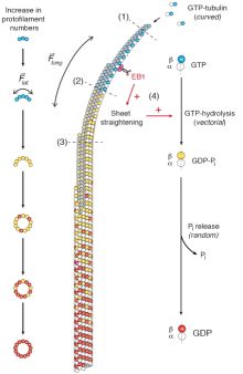 September 2016. Using gold nanoparticles functionalized with the multivalent chelator Ni-trisNTA, the microtubule plus-end tracking protein EB1 was site-specificly immobilized to investigate the architecture of the GTP-cap of growing microtubule ends by cryo-electron tomography. In a large collaborative effort, teams from France, Switzerland, Spain and Frankfurt provide unique structural information on the interaction of EB1 with growing microtubule ends. The study, published on 12 September 2016 by the journal Nature Cell Biology also offers insights into the conformational changes that tubulin dimers undergo during microtubule assembly and on the architecture of the GTP-cap region. More ...
Contacts:
Robert Tampé (tampe@em.uni-frankfurt.de) and Ralph Wieneke (wieneke@em.uni-frankfurt.de), Institute of Biochemistry, Riedberg Campus, Goethe-University Frankfurt, Frankfurt am Main, Germany
Publication:
Guesdon A, Bazile F, Buey RM, Mohan R, Monier S, Rodríguez García R, Angevin M, Heichette C, Wieneke R, Tampé R, Duchesne L, Akhmanova A, Steinmetz MO, Chrétien D (2016). EB1 interacts with outwardly curved and straight regions of the microtubule lattice. Nature Cell Biology, advanced online publication on 12 September 2016. http://dx.doi.org/10.1038/ncb3412